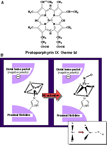
Figure 3. A: The structure of protoporphyrin IX (heme b) found in soluble guanylate cyclase. B: The current proposed model for NO-heme binding in soluble guanylate cyclase. The binding of NO to the ferrous iron results in the displacement of the proximal histidine from the heme iron. The inset is a side view parallel to the plane of the heme in order to emphasize the displacement of the ferrous iron from the plane of the porphyrin ring.